- Home
- Ultrasound: From Pregnancy to Musculoskeletal Imaging
Ultrasound: From Pregnancy to Musculoskeletal Imaging
- March 13, 2026
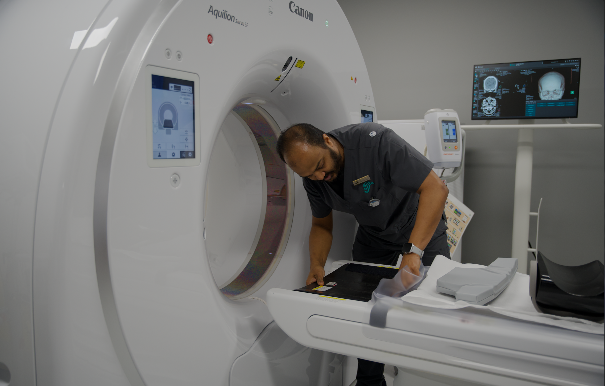
Ultrasound, also known as sonography, is a safe, non-invasive medical imaging technique that uses high-frequency sound waves to create real-time images of structures inside your body. Unlike X-rays or CT scans, ultrasound does not use ionizing radiation, making it a particularly safe option for many patients, including pregnant women and children. At Scope Radiology in Clayton, Victoria, we offer a comprehensive range of ultrasound services, performed by highly skilled sonographers and interpreted by Radiologists, to assist in accurate diagnosis across various medical specialties.
How Does Ultrasound Work?
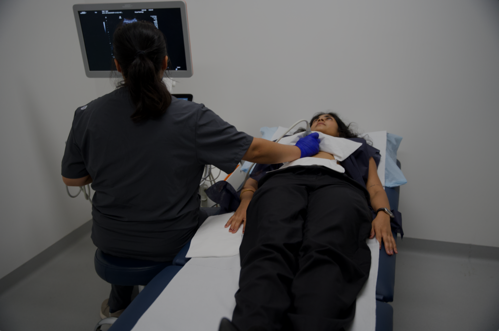
An ultrasound machine consists of a transducer (a small, handheld device) and a computer. The sonographer applies a special gel to the skin over the area to be examined. This gel helps the transducer make full contact with the skin and eliminates air pockets that could block the sound waves. The transducer then emits sound waves that travel into the body, bounce off organs and structures, and return to the transducer. The computer processes these reflected sound waves into live, moving images that can be viewed on a monitor.
Versatile Applications of Ultrasound
Ultrasound is a remarkably versatile diagnostic tool, used for a wide array of conditions and examinations:
1. Obstetric Ultrasound (Pregnancy Scans)
- Early Pregnancy: Confirming pregnancy, estimating gestational age, and checking for viability.
- Nuchal Translucency (NT) Scan: Performed in the first trimester to assess the risk of chromosomal abnormalities.
- Morphology Scan: A detailed examination around 18-20 weeks to assess fetal development, organ formation, and detect potential anomalies.
- Growth Scans: Monitoring fetal growth, position, and well-being in later stages of pregnancy.
2. Abdominal Ultrasound
- Organs: Examining the liver, gallbladder, pancreas, kidneys, and spleen for conditions like gallstones, kidney stones, fatty liver, or cysts.
- Blood Vessels: Assessing blood flow and detecting blockages or aneurysms.
3. Pelvic Ultrasound
- Female Pelvis: Evaluating the uterus, ovaries, and fallopian tubes for conditions such as fibroids, ovarian cysts, endometriosis, or causes of pelvic pain and abnormal bleeding.
- Male Pelvis: Examining the prostate and bladder.
4. Musculoskeletal (MSK) Ultrasound
- Joints, Muscles, Tendons, Ligaments: Diagnosing tears, inflammation (e.g., tendonitis, bursitis), sprains, and other injuries in areas like the shoulder, knee, ankle, and wrist.
- Soft Tissue Masses: Identifying lumps and bumps.
5. Vascular Ultrasound
- Blood Vessels: Assessing blood flow in arteries and veins, detecting deep vein thrombosis (DVT), arterial narrowing, or aneurysms.
6. Small Parts Ultrasound
- Thyroid: Examining the thyroid gland for nodules or goiter.
- Breast: Investigating breast lumps or abnormalities found on mammograms.
- Scrotum: Diagnosing conditions affecting the testicles.
Preparing for Your Ultrasound
Preparation for an ultrasound varies significantly depending on the type of scan. Our team will provide you with specific instructions, but common requirements include:
- Fasting: For abdominal ultrasounds (e.g., gallbladder, liver), you may need to fast for several hours beforehand.
- Full Bladder: For pelvic ultrasounds, you may be asked to drink water and avoid emptying your bladder to Aim to provide optimal visualization of pelvic organs.
- Comfortable Clothing: Wear loose, comfortable clothing. You may be asked to expose the area being examined.
- Referral: Always bring your valid doctor’s referral to your appointment.
What to Expect During the Procedure
When you arrive at Scope Radiology Clayton for your ultrasound:
- Check-in: Our reception staff will confirm your details and referral.
- Preparation: A qualified sonographer will explain the procedure and position you comfortably on an examination bed. Gel will be applied to your skin.
- The Scan: The sonographer will move the transducer over the area of interest, capturing images. You may feel slight pressure, but it should not be painful. You can watch the images on a screen if you wish.
- Duration: Most ultrasound scans take between 15 to 45 minutes, depending on the complexity of the examination.
Your Results
After your ultrasound, our Radiologists will meticulously review the images and the sonographer’s findings. A detailed report will then be sent to your referring doctor, typically within 24-48 hours. Your doctor will discuss the results with you and explain what they mean for your health and any necessary next steps.
At Scope Radiology in Clayton, we are committed to providing safe, accurate, and comprehensive ultrasound services. Our advanced technology and expert team Aim to provide you receive the highest standard of diagnostic care, from routine pregnancy scans to specialized musculoskeletal imaging.
Frequently Asked Questions (FAQ)
Yes, ultrasound is very safe. It uses high-frequency sound waves, not ionizing radiation, making it a preferred imaging method for many conditions, especially during pregnancy.
Preparation varies by the type of ultrasound. For example, abdominal ultrasounds often require fasting, while pelvic ultrasounds may require a full bladder. You will receive specific instructions when you book your appointment.
Most ultrasound examinations take between 15 to 45 minutes, depending on the complexity of the area being examined.
While the sonographer performs the scan, the images are then reviewed and formally reported by a Radiologist. Your referring doctor will receive the detailed report, typically within 24-48 hours, and will discuss the findings with you.
Musculoskeletal (MSK) ultrasound is used to diagnose conditions affecting muscles, tendons, ligaments, and joints. It’s excellent for detecting tears, inflammation (like tendonitis or bursitis), sprains, and other soft tissue injuries in real-time.